New material developed in a joint study between the Technion and the University of Chicago will pave the way for the restoration of damaged nerve tissue and heart muscle. Assistant Professor Hemi Rotenberg of the Faculty of Biomedical Engineering at the Technion and Professor Bozhi Tian of the University of Chicago have developed a way to project near-infrared light into a membrane of the new material, which will photo-activate the damaged nerve or heart tissue.
Nerve tissues are the biological platform that transmits information between different areas of the body. Most of them are found in the two control centers of the central nervous system: the brain and spinal cord. The peripheral nervous system stems from the central nervous system, controlling many physical activities, including muscle activation as well as the transmission of sensory information.
Damage to the peripheral nervous system can lead to limitations, such as paralysis, numbness, and chronic pain. Although peripheral nerves can undergo regeneration, the process is slow and the outcomes can be limited. Some medical interventions, such as electrical stimulation, may enhance the rehabilitation process. However, this method usually requires an invasive procedure that can damage the body’s tissues. This innovation may eliminate the need for electrode transplantation altogether.
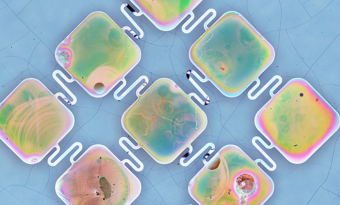
The researchers created a new, semiconductor device in a flexible, ultra-thin membrane configuration that interfaces well with biological tissues. They then used the membrane to wrap the damaged nerve tissue, or in the case of heart pacing, wrap the heart itself.
 “Our development is a photovoltaic material, that is, material that converts light energy into electrical energy that affects nerve tissue,” explained Prof. Rotenberg. “We have demonstrated the efficacy of the new substance in two different contexts — heart pacing and the activation of the peripheral nervous system.”
“In the context of heart treatments, for example, the use of such a device can allow temporary cardiac pacing for post-operative rehabilitation and avoid the use of a temporary electrode to be inserted into the heart. Because the membrane we developed is made of a silicon-based material, which absorbs in the body without any toxic effect whatsoever, there is no need for further surgical action to remove it from the body.”
The uniqueness of the material lies in the formation of a semiconducting diode junction from a single type of silicon. This is highly unusual, as usually diodes are made by interfacing two types of silicon. Semiconductors are based on energetic gaps that determine their level of conductivity; they are usually made up of n-type materials, which contribute an electron to the material, and p-type materials that take an electron from the material (leaving a hole instead); the connection between the two materials creates an efficient interface called p-n junction, which is the building block of electronic devices and solar cells.
According to Prof. Rotenberg, the new material was created in an unplanned way. “I accidentally used a metal tweezer in the laboratory, which provided iron ions to the solution — something I did not plan to happen. The iron ions turned out to catalyze the creation of nano-pores on the surface of silicon.” He also says the new material is a therapeutic window that allows the medical team to have an external impact on the tissues of the patient’s body.
Outside the medical field, the new development is expected to contribute greatly to various applications, for example, in the field of renewable energies. Since renewable energy sources such as the sun are volatile, and do not operate at constant intensity throughout the day, energy storage becomes a major challenge in promoting the use of these energies. Prof. Rotenberg estimates and hopes that the new material he developed with his colleagues will accelerate the development of more advanced and efficient solar devices.